DUAL HEAD SPECT

ECAM NOVA HIGHLIGHTS

Advanced acquisition software (MIA)
Improved user interface
Linked protocols
Auto multi-position SPECT
Multi-tracer protocols
Online reconstruction

touchscreen
Integrated touchscreen for convenient patient positioning and camera operation
PPM allows to trigger automatic detector movements for a fast work routine

FRED 59 DIGITAL Detector
HD digital detectors with consistent image quality
59 PMTs per detector for high resolution
High sensitivity for lower dose and faster scans
Lead shielding up to 511 keV
3/8″ NaI(Tl) crystal
Real-time body contouring for close positioning

Flexible detector configurations
76°, 90° & 180°
76° configuration optimized for myocardial SPECT imaging
Closer detector positioning along the patient’s chest
Beneficial for slim patients and prone imaging
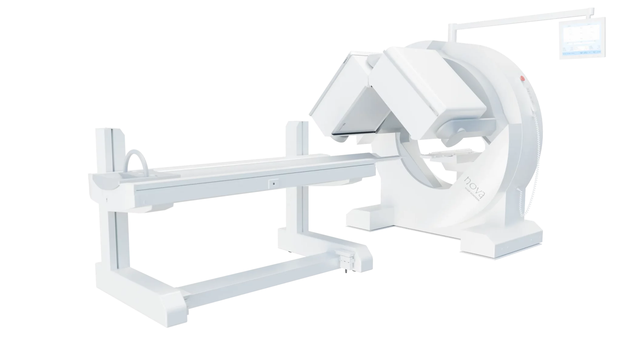
Meet ecam nova
Gamma camera for SPECT, whole-body, and planar imaging designed for high patient throughput
from 20° cranial to 90° caudal

ensure high patient comfort - no additional parts required

Delivering excellent image quality with energy-independent performance
High sensitivity enabling optimized dose and reduced acquisition time

for convenient patient positioning and camera operation

for excellent resolution

76°, 90°, and 180°
The 76° configuration is optimized for myocardial SPECT imaging. It allows the detectors to follow the patient’s chest more closely during acquisition, which can be particularly beneficial for slim patients or during prone imaging.

for easy access for patients with limited mobility (48.3 cm / 19”)


SCINTRON 7 NM
Software Highlights
The SCINTRON software contains a wide variety of organ specific processing programs. In addition, general processing tools allow for evaluation of studies customized to your needs. Quantification and viewing made easy by intuitive program structure with fast access of result screens. Individual masks and export functions round out the results to fit your clinical and documentation requirements. The SCINTRON system includes a DICOM-interface for seamless communication with RIS/HIS and PACS. The workstation allows for parallel acquisition and processing for a time saving work routine.

SPECT Software
- 3D iterative reconstruction (DROSEM) for highest reconstructed spatial resolution
- Flexible slice displays for individual screen layouts
- Assembling of up to five SPECT acquired in multiple bed positions

Lung
- Calculation and displaying of V/P-Quotient in the slice viewer
- Direct comparison of ventilation and perfusion studies

whole body
- Dual-intensity display to evaluate posterior and anterior views
- Quantification of sacroiliac joints within whole body scan, no additional static image required
- Deconvolution filter for improvement of signal / noise ratio

MyoCard
- Display of rest / stress bullseye plots and reversibility
- Assessment of EF, wall thickening and wall motion for gated SPECT
- Calculation of SSS, SRS, and SDS with the use of an individual normal database
- Washout calculations for thallium studies
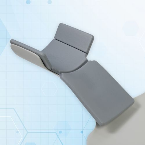
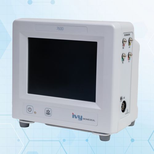
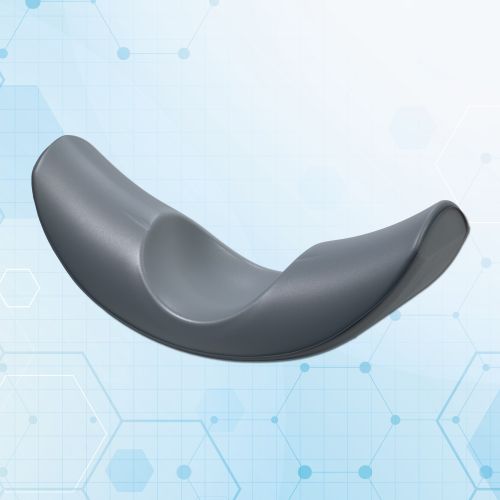
Monitor & patient positioning
Technical Details
|
|
|
| 53.3 cm x 38.7 cm (21” x 5.25”) | |
| Number of PMTs | |
| Crystal Thickness | |
| Energy Range | |
| System sensitivity (with LEHR-Collimator) |
| Positioning | |
| Detector Tilt Range | |
| Detector Rotation Range | |
| Average Autocontour Distance | |
|
Detector configurations
|
|
|
|
| 202 cm (79.5”) | |
| Vertical Motion Range |
|
|
|
| Dual Detector 1580 kg (3485 lbs) |
|
|
|
| MEGP | |
| Pinhole | |
| High Energy |
|
|
|
| Brain SPECT | |
| ECG Trigger | |
| Additional Phantoms for Quality Control available |